In this article8
- What robotic graft selection means in hair transplantation
- What robotic graft selection can assess from images
- Why visible features do not equal graft viability
- Robotic graft selection cannot replace donor-area judgement
- Hairline design and placement remain human decisions
- Questions to ask when a clinic mentions automation
- How technology fits into a safe treatment pathway
- Frequently Asked Questions
Robotic graft selection can help identify and sort follicular units using measurable visual features, such as hair number, size, angle, and image-based appearance. It cannot independently judge your full donor safety, future hair-loss pattern, scalp health, or the artistic choices needed for a natural hairline. A qualified doctor should review every treatment decision.
- Automated systems can support follicular-unit assessment, but they do not replace clinical examination.
- Hair count, unit size, direction, and visible image features are useful data points, not complete measures of graft quality.
- Donor-area planning depends on long-term hair-loss risk, scalp condition, and safe extraction limits.
- A natural-looking result requires medical judgement and artistic placement, especially at the hairline.
- Questions about bone grafts are medically separate from hair transplantation and should be discussed with the appropriate specialist.
01What robotic graft selection means in hair transplantation
Robotic graft selection describes the use of computer-guided imaging and software to assist with identifying, assessing, or sorting follicular units during hair transplantation. A follicular unit is a naturally occurring group of one to several hairs that emerges from the scalp. In the right clinical setting, technology may make some parts of graft assessment more consistent and may help the surgical team organise information. However, it is important to understand that a machine-based assessment is only one part of the process. It does not turn hair transplantation into an automatic procedure, and it does not remove the need for an experienced medical team.
The term can be used differently by different providers. Sometimes it refers to imaging that maps the donor area before extraction. In other situations, it refers to software that classifies grafts after they have been removed, for example by the apparent number of hairs in each unit. It may also describe robotic assistance with donor-area scanning or extraction. These are related but different stages, and patients should ask clearly which stage the technology supports. The answer helps set realistic expectations about what is automated and what remains dependent on human assessment.
Hair graft evaluation is more complex than simply finding the units with the most hairs. Each graft needs to be considered in relation to where it may be placed, how it was handled, whether it appears intact, and whether its use supports a balanced long-term plan. Fine single-hair units can be especially valuable near a frontal hairline, while multi-hair units may be more suitable for selected areas behind it. The final distribution should be based on facial proportions, existing hair, donor capacity, and the patient’s likely pattern of ongoing hair loss.
02What robotic graft selection can assess from images
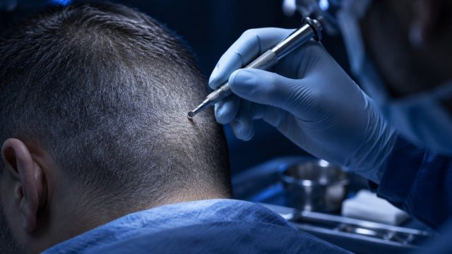
When image quality is good, a system may be able to measure visible characteristics of follicular units with useful speed. These can include the approximate number of hair shafts emerging from a unit, the spacing between visible follicles, shaft thickness as it appears in the image, and the angle at which hairs leave the scalp. It may also help identify areas that look denser or less dense. This information can support documentation and give the team a more structured view of the donor region before or during a procedure.
Automated image analysis may be particularly helpful when there are many grafts or a large donor area to review. It can reduce repetitive counting tasks and can flag units for closer inspection. For example, if a graft appears to contain several hairs, software may classify it differently from a finer unit with one visible hair. Used carefully, this can assist workflow and may help maintain an orderly approach to graft grouping. The output should still be checked because hair overlap, lighting, moisture, trimming length, and camera angle can affect what an image appears to show.
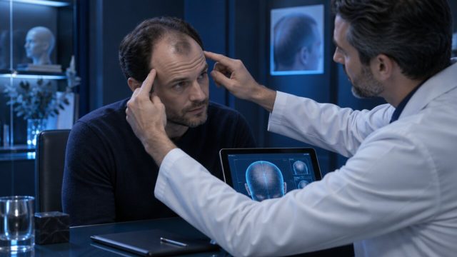
Some systems can also create a visual donor map. This may help record where extractions have taken place and may assist the team in spreading removals across an appropriate zone. A well-distributed pattern matters because taking too many grafts from a limited area can leave the donor region looking thinner. Mapping is informative, but it is not a substitute for the doctor’s direct examination. The scalp must be assessed in person or through a suitable medical consultation, including consideration of hair calibre, curl, contrast between hair and scalp, and signs of miniaturisation.
03Why visible features do not equal graft viability
A graft can look promising in an image and still require careful handling and professional review. Hair count is not the same as graft survival. A camera cannot fully confirm whether delicate tissue around the follicles has been affected during extraction, whether the graft has experienced excessive drying, or whether its storage conditions have been appropriate. These factors can influence how well a graft performs after implantation. For this reason, visual classification should be seen as supportive information rather than a guarantee of biological quality.
Follicular units are living tissue. Their condition depends on more than the number of shafts that can be counted. The surrounding tissue, the way the graft was dissected or extracted, the time it spends outside the body, hydration, temperature, and placement technique are all clinically relevant. An experienced team monitors these practical details throughout the procedure. Software may help organise graft categories, but it cannot independently manage tissue handling or recognise every concern that an experienced clinician may notice under magnification.
It is also not possible to predict an exact cosmetic outcome by looking at a graft alone. Growth depends on healing, blood supply in the recipient area, scalp characteristics, post-procedure care, and individual biological variation. In most patients, visible changes develop gradually over months rather than immediately. At Acibadem Hair Transplant Center, results are measured by trichoscopic graft survival at month 12, with a documented 98% rate under written guarantee. Your doctor will explain how outcome assessment applies to your treatment plan and why individual results can vary.
04Robotic graft selection cannot replace donor-area judgement
The donor area is a limited resource, usually located at the back and sides of the scalp. A key medical decision is determining how many grafts may be removed while preserving an acceptable donor appearance. This decision cannot be made safely from a simple count of visible follicular units. The doctor must assess scalp laxity where relevant, distribution of density, hair thickness, possible miniaturisation, prior procedures, scars, and the likelihood that hair loss may progress in other areas.
Robotic graft selection may identify units that appear technically suitable for extraction, but it cannot independently determine the best long-term extraction pattern for every patient. For example, a donor area may look dense in one small zone while being less uniform across the wider safe region. Removing repeatedly from the densest-looking section could create an uneven appearance. A clinician considers the whole donor landscape and plans extraction distribution accordingly. This is particularly important for patients who may need to preserve grafts for a future procedure.
A doctor also needs to distinguish stable donor hair from hairs that may be vulnerable to thinning. This requires medical judgement based on examination, history, family pattern, and, when appropriate, trichoscopic assessment. Algorithms can recognise patterns in images, but they do not replace an evaluation of why a pattern is present or how it may change over time. A responsible plan should prioritise donor preservation and realistic coverage rather than pursue the highest possible number of extracted units.
05Hairline design and placement remain human decisions
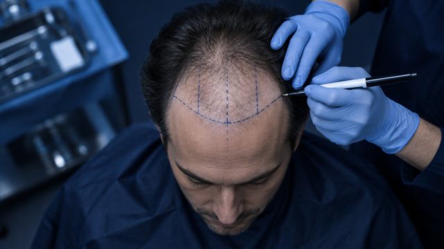
Natural hair restoration is not only about placing a certain number of grafts. It involves deciding where each category of graft is most appropriate. The frontal hairline usually needs softness, irregularity, and fine detail so that it blends naturally with existing hair. Temple points, frontal corners, and transition zones also require close attention to direction and angle. A system can sort units by visible features, but it cannot fully understand the aesthetic effect that placement will have on an individual face.
Doctors and trained implantation teams consider facial balance, age, ethnic features, current hairline position, and the possibility of future loss when creating a plan. A very low or overly dense hairline may not remain appropriate if thinning progresses. Similarly, the direction of implanted hairs needs to follow a pattern that looks natural in everyday movement, not only in a single image. These decisions are based on training, observation, and an understanding of how transplanted and native hair will work together.
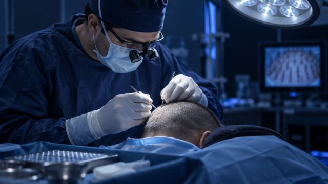
Graft placement also requires gentle, precise technique. The recipient sites must be made at suitable angles, depths, and spacing to support growth and avoid an unnatural appearance. Image analysis cannot assess the tactile feedback of the scalp or make real-time adjustments when tissue characteristics vary. Whether a patient has Sapphire FUE or DHI, the clinical team must make careful decisions throughout the procedure. Technology can be useful, but the treatment should remain medically led and individually planned.
06Questions to ask when a clinic mentions automation
If a clinic mentions robotic graft selection, it is reasonable to ask what the technology actually does. Does it only scan the donor area, identify follicular units, assist with extraction, sort grafts after removal, or create a density map? You can also ask who verifies the system’s findings and who makes the final decisions about extraction and implantation. Clear answers are a positive sign because they show that technology is being presented as a tool rather than as a replacement for clinical responsibility.
It is helpful to ask how the donor area will be protected, how grafts are stored and handled, and how the team plans graft placement for your goals. You may also ask how the clinic assesses progress after the procedure. At Acibadem Hair Transplant Center in Istanbul, patients can request a free online hair analysis that is reviewed by doctors, with WhatsApp replies typically available within around two hours. This initial review can explain whether a fuller consultation is appropriate, but it does not replace a complete medical assessment.
Patients should be cautious about claims that any device can guarantee a certain density, exact growth rate, or a completely painless experience. Needle-free anaesthesia may be available for suitable patients, yet your doctor will confirm whether it is appropriate. A clinic licensed by the Turkish Ministry of Health should provide a clear explanation of the process, risks, aftercare, and expected timeline. A well-informed decision is usually based on the team’s medical standards, transparency, and follow-up plan rather than on the name of a single machine.
07How technology fits into a safe treatment pathway
Technology is most useful when it supports a structured clinical pathway. Before a procedure, imaging and trichoscopic review can help document the condition of the hair and scalp. During the procedure, magnification, organised graft handling, and careful recording can support precision. After the procedure, follow-up photographs and examinations can help monitor progress over time. Each step provides information, but the information needs medical interpretation. No device can fully account for your medical history, medications, healing pattern, or personal expectations without clinician involvement.
For international patients, practical support can also make it easier to focus on recovery. Acibadem Hair Transplant Center is a dedicated hair restoration clinic in Istanbul, licensed by the Turkish Ministry of Health, and has welcomed patients from 38 countries over 12 years. Its all-inclusive packages include VIP transfer, a 4-star hotel, a care kit, and 12 months of follow-up. These services are designed to support the patient journey, while the suitability of any procedure remains a medical decision confirmed by the doctor.
In summary, automated graft assessment may add useful information about visible follicular-unit features and help a team work efficiently. Its limits are equally important: it cannot independently judge graft viability, protect the donor area, predict every future change, or create an aesthetically appropriate hairline. The most reliable approach combines suitable technology with direct medical examination, careful surgical technique, and long-term planning. Before choosing treatment, ask how clinical judgement guides every stage from donor analysis to follow-up.
08Frequently Asked Questions
What are the four types of grafts?
In bone grafting, the four broad categories are autografts, allografts, xenografts, and synthetic graft materials. An autograft comes from the same person, an allograft comes from another human donor, a xenograft comes from an animal source, and a synthetic graft is manufactured. These categories are separate from hair transplant grafts, which are follicular units containing one or more hairs.
What type of bone graft is best?
There is no single best bone graft for every situation. An autograft is often considered a valuable option because it uses the patient’s own tissue, but it may require a second surgical site and is not suitable in all cases. The best choice depends on the bone location, size of the defect, health factors, surgical goals, and the specialist’s assessment.
How long does a graft take to heal?
For bone grafting, initial healing can take several weeks, while integration and remodelling may take several months or longer depending on the procedure and the person’s health. Your surgeon will use examinations and, where needed, imaging to assess healing. In hair transplantation, implanted follicles also settle gradually, and visible hair growth typically develops over months rather than days or weeks.
What are the downsides of allografts?
In bone grafting, allografts may have slower biological incorporation than an autograft because they do not contain the patient’s own living cells. They also have limited osteogenic potential after processing, and although donor screening and processing are designed to reduce risk, patients may have concerns about disease transmission or immune response. Suitability should be discussed with the surgeon managing the bone condition; this topic does not apply to follicular-unit grafts used in hair transplantation.
This article is for general information only and is not a substitute for professional medical advice.
This guide is for general information and does not replace a medical consultation. Treatment decisions are made after an in-person or online assessment by a doctor.